Radiology
Radiology Services
We offer a variety of diagnostic imaging studies onsite. The images are transmitted to and interpreted by radiologists in Bismarck, ND, and patients receive same-day results.
- X-rays: We perform x-rays of the chest, abdomen, spine, extremities, and other bone or soft tissue areas of the body.
- CT Scan: CT scans provide more detailed information than regular x-rays by combining a series of x-ray images of the body as well as the bones, blood vessels, and soft tissue.
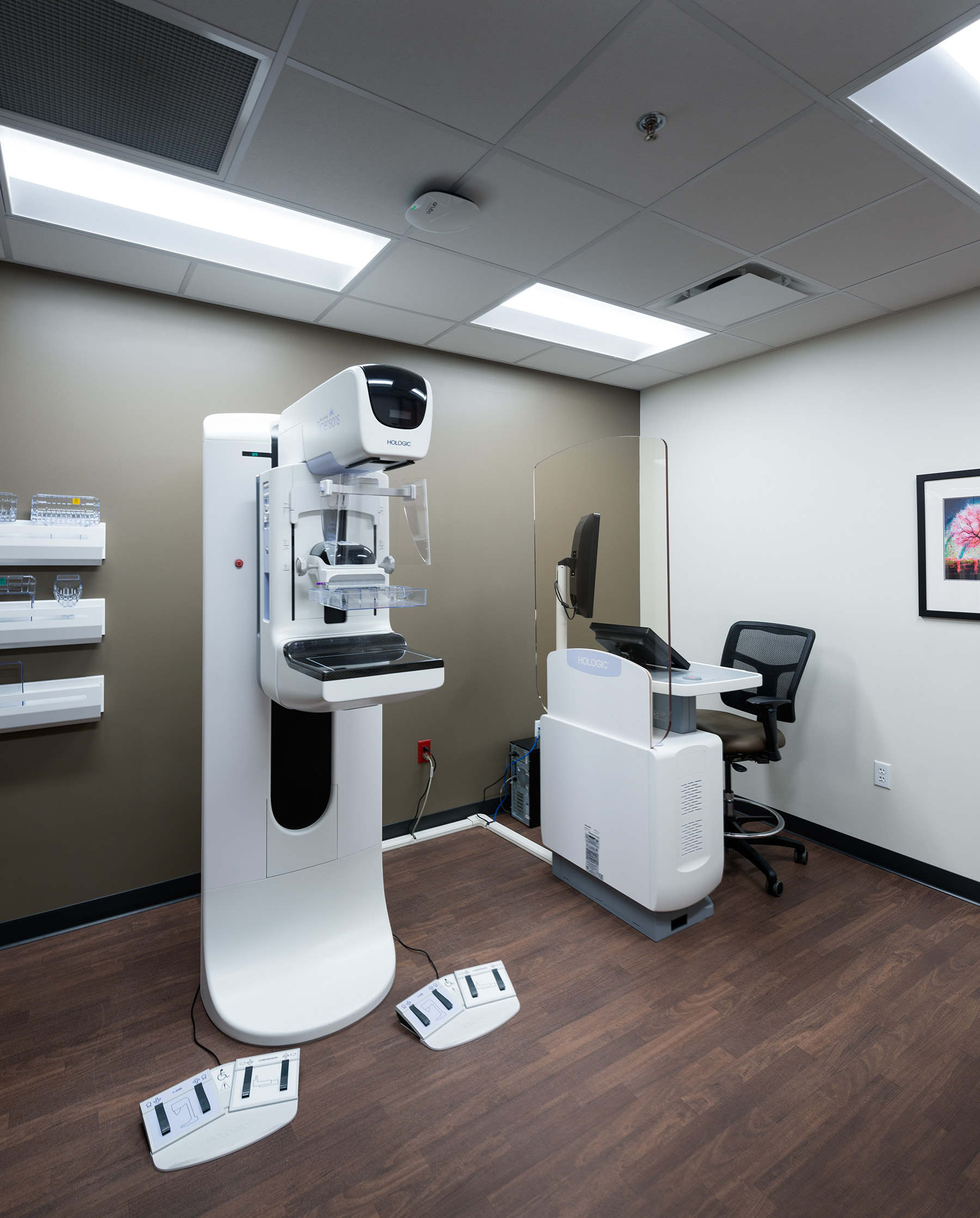
- Mammograms: We offer 3D mammography which helps identify individual breast structures without the confusion of overlapping tissue.
- Ultrasound: Diagnostic ultrasounds use sound waves to produce images of your body. They're commonly used during pregnancy to monitor the fetus, but can also be used to image the thyroid, abdominal organs, and much more.
- Bone Densitometry (DEXA Scan): This procedure measures bone mass which can help determine risk of fractures and/or osteoporosis.
- MRI:* Without using traditional x-ray imaging, Magnetic Resonance Imaging (MRI) allows doctors to see inside the body. An MRI samples signals from the water that makes up your body.
- Echocardiogram:* An echocardiogram uses sound waves to show how blood flows through the heart and heart valves. This test can help diagnose heart conditions.
- Nuclear Medicine Exam:* Nuclear medicine exams use small amounts of radioactive material to diagnose many diseases like cancer, heart disease, and other abnormalities within the body.
We work with a number of external partners to offer specialized imaging options at our facility. An asterisk* indicates services offered regularly onsite through mobile service.
Hours & Contact
SMC Radiology
(701) 748-7278
Monday - Friday
7:00 a.m. to 5:00 p.m.
Our staff is on-call 24/7 in case of emergency.
Our Equipment
We proudly offer the latest advanced digital imaging technology to help diagnose and treat a variety of medical conditions. Our state-of-the-art equipment is completely digital and filmless. Studies are stored electronically which allows them to be sent almost anywhere in North Dakota.
When you need a scan, reach out to our radiology department. Our wait time is typically much shorter than large facilities.
.jpg)